
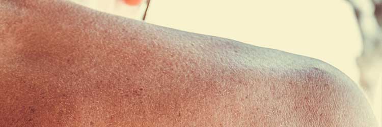
1. Skin Care
Tissue viability care is required whenever there is a potential or actual risk to tissue health.
Tissue health is affected by both irreversible and reversible factors - therefore, reducing these threats promotes tissue health.
One irreversible factor is the ageing process. Reversible factors include poor perfusion, oxygenation and nutrition (Mallet 2013).
The first section of this article will briefly focus on reducing reversible factors that can cause pressure injuries.
Pressure Injury
A pressure injury is defined as a ‘localised injury to the skin and/or underlying tissue, usually over a bony prominence, as a result of pressure, or pressure in combination with shear and/or friction’ (ACSQHC 2018). All critically ill patients are at especially high risk and therefore require pressure area care.
Factors that contribute to pressure injury include:
- Pressure - any intense or prolonged pressure, usually from body weight, deprives skin and soft tissue cells of oxygen and nutrients.
- Shear force - caused by a force parallel to the surface of the skin.
- Friction - caused by two surfaces rubbing against each other, possibly due to poor moving and handling techniques. Friction injuries can also cause abrasions, superficial ulceration and blistering.
(EPUAP/NPIAP/PPPIA 2019)
Risk factors for pressure injury include:
- Malnutrition or dehydration
- Inability to reposition self
- Reduced mobility
- Loss of sensation
- Comorbidities (e.g. neurologic disease, cardiovascular disease, anaemia)
- Reduced skin moisture
- Incontinence
- Older age
- Cognitive impairment
- Surgical patient
- Prolonged anaesthesia.
(Zaidi & Sharma 2024)
Pressure Injury Risk Assessment
Pressure care management is essential in critically ill patients, as they generally have several risk factors present. Consider using a validated risk assessment tool to support your clinical judgement. Any indication of risk should be followed by action.
Performing a Pressure Injury Risk Assessment
Risk assessment should be undertaken within 8 hours of admission to the hospital (Vic DoH 2024).
- Examine the patient’s entire skin for skin damage by looking at and touching the skin from head to toe, with an emphasis on bony prominences and skin folds.
- Reassess pressure injury risk if there is a change in the patient’s clinical condition such as deterioration or surgery.
- The patient’s skin should be inspected on an ongoing basis, depending on the clinical setting and the patient’s degree of risk.
- The patient’s skin should be examined before the patient is discharged.
- Always follow your local policies and recommendations for risk assessment, documentation and how to communicate information to the interprofessional team.
(Mallet 2013)
Every skin assessment should include any pain or discomfort reported by the patient. Check for:
- Skin temperature
- Dryness
- Oedema
- Colour changes or discolouration
- Bruising
- Inflammation
- Scratch marks
- Jaundice
- Swelling
- Breaks in the skin
- Ulcers
- Lesions
- Rashes.
(Vic DoH 2024)
Pressure Injury Prevention
Risk factors can be reduced by:
- Optimising hydration and nutrition
- Managing incontinence
- Changing a patient’s clothes and linen if they become moist
- Performing routine skin care
- Encouraging mobilisation
- Reviewing the patient’s medications
- Frequent repositioning
- Using pressure-redistributing equipment.
(Vic DoH 2024)
Any signs of actual or potential risks should be assessed and documented, and appropriate action taken to promote healing and prevent further skin damage.
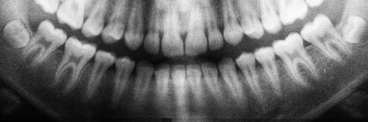
2. Mouth Care
Oral care is an essential nursing activity that provides relief and comfort to patients who are seriously ill and cannot perform simple activities of daily living themselves.
The oral cavity and other parts of the mouth are perfect media in which bacteria can live and thrive. Therefore, all parts of the oral cavity should be assessed each time the mouth is cleaned (Mallet 2013).
Assessment of the Oral Cavity
- Follow any oral assessment tool available within your organisation to ensure consistency and facilitate a thorough examination.
- Ensure you have sufficient light to visualise as much of the oral cavity as possible.
- Ensure the patient’s head is appropriately supported (e.g. with pillows) to prevent trauma or discomfort.
- Ensure linen is not touching the patient’s lips as linen can cause drying and discomfort.
- Document care and any abnormalities to provide an accurate record, monitor the effectiveness of the procedure, and facilitate communication and continuity of care.
(Mallet 2013)
Oral Cavity Care
- Lips: Assess for ulceration or ‘cracking’, which is mainly caused by dryness, dehumidified oxygen via facemasks, or damage from endotracheal tape/holder. Provided there is no sign of infection, apply a lubricant such as soft yellow paraffin, humidify face mask oxygen, and, if possible, prevent the tape securing the endotracheal tube from touching the patient’s lips (especially at the corners of the mouth). If not possible, change the position of tapes on lips at least once daily and use any available aids to reduce trauma.
- Teeth: Assess for damaged or broken teeth caused by trauma or poor oral hygiene. Sit the patient upright (unless contraindicated) and clean their teeth with a toothbrush, usually twice daily.
- Gums: Assess for bleeding caused by trauma or poor hygiene. Clean gums when cleaning teeth.
- Saliva: Assess for excess saliva, lack of high viscosity (possibly caused by objects in the mouth such as an endotracheal tube) or dehydration. Moisten mouth if dry and increase the frequency of moistening mouth.
- Tongue: Assess for dark colour and dryness, possibly caused by reduced perfusion and dehydration.
- If the patient can clean their own teeth, they should be encouraged to do so.
(Mallet 2013)
Denture Hygiene
- Remove dentures from an unconscious patient to prevent airway obstruction
- Remove dentures overnight from patients who are conscious in order to facilitate cleaning
- Brush dentures with a toothbrush to remove debris
- Rinse dentures under running cold water and dry thoroughly. Dentures can be damaged if left dry or cleaned in hot water
- If not worn by the patient during the day, store dentures safely in a labelled denture pot within the patient’s own property.
(Clarke 1993; Mallet 2013)
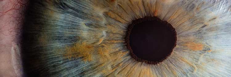
3. Eye Care
Vision is one of the main senses and a means of communication for most people. Impaired vision can, therefore, contribute to delirium.
Ocular diseases rarely require critical care admission, but pre-existing conditions may need continuing treatment, such as eye drops (Dawson 2005).
Eye care refers to measures that maintain ocular health and comfort. In critical care, this usually means care given to protect eye surfaces from potential harm, treatments for specific (acute or chronic) problems, and care of visual aids such as glasses and contact lenses (Mallet 2013).
The aim of eye care is to:
- Prevent potential harm/trauma
- Treat any identified problems
- Care for and clean any visual aids.
Factors that may expose eyes to potential damage in critical care include:
- Altered state of consciousness
- Metabolic derangement
- Immunocompromise
- Mechanical ventilation
- Certain medications (e.g. sedatives, muscle relaxants)
- Paralysis
- Exposure to respiratory pathogens
- Systemic disease
- Being in the prone position.
(ACI 2024)
Suspected ocular infections should be recorded and communicated to relevant staff members.
Eye Assessment
Eye health assessments should be part of a routine patient physical assessment and be performed on admission, followed by an ongoing assessment at the beginning of each new nursing shift.
Assessment should include:
- Using a bright light to examine the eyes, looking for signs of infection or disease such as:
- Redness, pain or discharge
- Swelling of the eyelids and conjunctiva
- Lid margin crusting
- Corneal clouding
- Following any eye assessment tools used within your organisation
- Ensuring the patient’s head is supported at a sufficient angle to prevent periorbital oedema and intraocular hypertension
- Ensuring linen (especially seams) and equipment are not in direct contact with either eye. Anything touching the eye surface can cause trauma
- Visualising both eyes for assessment
- If the patient normally uses visual aids (glasses or contact lenses), this should be documented, whether aids are present or absent, in use or not
- Visual aids should only be used if the patient wishes to and if they are conscious and in a suitable position (upright).
(ACI 2024; Cooke et al. 2011; Mallet 2013)
Test Your Knowledge
Question 1 of 3
When should a pressure injury risk assessment be performed?
Topics
References
- Agency for Clinical Innovation 2024, Eye Care of the Critically Ill, New South Wales Government, viewed 18 September 2024, https://aci.health.nsw.gov.au/networks/icnsw/clinicians/eye-care
- Australian Commission on Safety and Quality in Health Care 2018, Pressure Injury, Australian Government, viewed 17 September 2024, https://www.safetyandquality.gov.au/sites/default/files/2019-05/saq7730_hac_factsheet_pressureinjury_longv2.pdf
- Clarke, G 1993, ‘Mouthcare and the Hospitalised Patient’, British Journal of Nursing, vol. 2, no. 4, viewed 18 September 2024, https://www.magonlinelibrary.com/doi/abs/10.12968/bjon.1993.2.4.225
- Dawson, D 2005, ‘Development of a New Eye Care Guideline for Critically Ill Patients’, Intensive and Critical Care Nursing, vol. 21, no. 2, viewed 18 September 2024, https://www.sciencedirect.com/science/article/abs/pii/S0964339705000169?via%3Dihub
- Cooke, K, Doyle, N & Farley, A 2011, ‘Patient Comfort’, in Dougherty, L & Lister, S (eds), The Royal Marsden Hospital Manual of Clinical Nursing Procedures, 8th edition, Oxford: Wiley-Blackwell.
- European Pressure Ulcer Advisory Panel, National Pressure Injury Advisory Panel and Pan Pacific Pressure Injury Alliance 2019, Prevention and Treatment of Pressure Ulcers/Injuries: Clinical Practice Guideline: The International Guideline 2019, EPUAP NPIAP & PPPIA, viewed 18 September 2024, http://www.internationalguideline.com/static/pdfs/Quick_Reference_Guide-10Mar2019.pdf
- Mallet, J, Albarran, J & Richardson, R 2013, Critical Care Manual of Clinical Procedures and Competencies, Wiley-Blackwell, Oxford.
- Victoria Department of Health 2024, Identifying and Preventing Skin Problems, Victoria State Government, viewed 18 September 2024, https://www.health.vic.gov.au/older-people-in-hospital/pressure-injuries-and-skin-tears-0/identifying-and-preventing-skin-tears
- Zaidi, SRH & Sharma, S 2024, ‘Pressure Ulcer’, StatPearls, viewed 18 September 2024, https://www.ncbi.nlm.nih.gov/books/NBK553107/