Renal Function
Renal function is the key indicator of the kidneys’ condition and should be monitored in all deteriorating or critically ill patients in order to:
- Monitor disease progress
- Assess baseline measurements before starting treatment with certain medicines
- Indicate the function of other systems (e.g., cardiovascular system, where low cardiac output will result in diminished urine output)
- Identify renal impairment.
This article will discuss the principles of monitoring renal failure.
The Kidneys
The kidneys, located near the middle of the back, just below the rib cage, have several functions, which include:
- Secretion of hormones such as:
- Renin: Is vital for the activation of the renin-angiotensin-aldosterone system, which plays a vital role in increasing blood volume regulation of fluid balance
- Erythropoietin, which is synthesized in the kidney and stimulates the bone marrow to produce red blood cells
- Regulation and maintenance of:
- Fluid balance
- Electrolyte balance
- Acid-base balance
- Excretion of foreign materials such as medicines and by-products of metabolism, nitrogen, urea, and creatinine
- Maintenance of calcium and phosphorus balance, and activation of vitamin D.
(You and Your Hormones 2019; NIDDK 2018; Gounden et al. 2023)
Principles of Urinalysis
A urinalysis can provide healthcare professionals with valuable information about the patient’s health status. For example, urinalysis can provide indications of kidney disease, diabetes mellitus, liver disease, urinary tract infection (UTI), and general dehydration (Milani & Jialal 2023).
The Purpose of Urinalysis
- Screening for systematic diseases such as renal conditions and diabetes mellitus
- Diagnosis - to confirm and exclude suspected conditions, for example, UTIs
- Management and planning - to monitor the progress of an existing condition and plan programs of care.
(Milani & Jialal 2023)
Appearance of Urine
The colour of urine can vary greatly. Normal urine varies in appearance from clear to straw-coloured and is practically odourless but becomes turbid and smells of ammonia if left to stand (Rotker & Watson 2023; Labpedia.net 2023).

Variations in the appearance of urine include the following:
- Clear: Very clear and colourless urine may indicate excessive fluid intake. It may also be caused by pharmacologically induced diuresis or diabetes insipidus/diabetes mellitus
- Dark: Urine is concentrated as seen in fluid depletion or contains conjugated bilirubin and jaundice
- Orange: Can indicate dehydration, or can be caused by certain vitamins or medications
- Pink/red: May indicate haematuria, though other causes include ingestion of certain foodstuffs like beetroot and blackberries
- Cloudy: May indicate the presence of pus, protein or white blood cells and requires further investigation
- Sediment: Can indicate a range of conditions, such as urinary tract infection, diabetes and bladder stones
- Frothy: May indicate significant proteinuria.
(Chavoustie & Sissons 2023; Cadogan 2023; Haase 2022; Luo & Herndon 2023; Mayo Clinic 2023)
Urine Odour
- Normal, freshly voided urine is practically odourless. If left to stand for several hours, it acquires a mild smell of ammonia
- Infected urine has a ‘fishy’ smell
- In patients with diabetes who have ketoacidosis, acetone is excreted in the urine, causing the urine to smell uncharacteristically sweet. Acetone can also indicate starvation.
(Labpedia.net 2023; Biggers & Gotter 2018; ACT Health 2019)
Dipstick Urinalysis
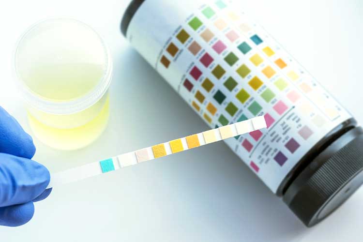
A dipstick urinalysis can accurately show the presence of a variety of substances such as protein, glucose, blood, and ketones as well as the PH.
To ensure reliable results:
- Always use a fresh sample of urine collected in a clean receptacle
- Observe the sample for colour, appearance, smell and debris
- Ensure that the reagent strip is in date
- Ensure that the whole of the reagent strip is immersed in the urine sample
- Tap the strip on the side to remove excess urine, place horizontal and compare reagent pads with the colour scale at time intervals stipulated by the manufacturer, documenting results immediately
- Safely discard the strip and urine sample
- Store reagent strips following the manufacturer’s recommendations.
(ACT Health 2019; Jevon 2012)
Significance of Urinalysis Results
- Glucose in urine indicates that blood glucose is raised and the consolidation of glucose in the plasma exceeds the renal threshold. Causes include diabetes mellitus, Cushing’s syndrome and stress
- Ketones are by-products of fat metabolism and are suggestive of excessive fat breakdown as in starvation, fasting and uncontrolled diabetes
- Proteinuria is the presence of abnormally large quantities of protein. Persistent proteinuria is typically a sign of renal disease
- Haematuria (presence of blood) is associated with diseases of the kidney or urinary tract; it can also be present during menstruation
- Nitrate is strongly indicative of infection, although negative results can’t rule out infection
- White blood cells (pyuria) usually indicate an infection somewhere along the urinary tract and are an indication for laboratory testing.
(ACT Health 2019; National Kidney Foundation 2022; Cleveland Clinic 2022)
Principles of Urine Output Monitoring
- Urine output is frequently used as a guide to the adequacy of cardiac output (renal perfusion amounts to 20 to 25% of the cardiac output)
- The average urine output in a healthy adult is 800 to 2,000 mL/day
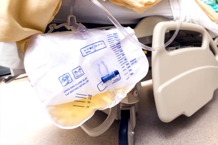
- All critically ill patients will require a urinary catheter to measure urine output and generally, an hourly urine drainage bag will be attached
- The urinary catheter should be closely monitored for blockages or occlusions
- If urine output dramatically falls, always consider mechanical obstruction first
- Sometimes bladder washouts are indicated (intermittent or continuous).
(Kaufman et al. 2023; MedlinePlus 2023; ACI 2022)

Principles of Monitoring Fluid Balance
Monitoring fluid balance in critical illness is essential. Having an understanding of disease processes is essential because clinical conditions can deteriorate rapidly.
- Careful monitoring of the fluid balance chart (input and output) must be maintained
- The patient should also be monitored for signs of fluid loss/gain
- Daily measurement of serum sodium, potassium, urea and creatinine is required to assess fluid and electrolyte balance
- Fluid balance charts from the preceding few days should be compared with serum and urine urea and electrolyte values to help evaluate the patient’s response to fluid administration and guide the fluid regimen over the next 12 to 24 hours.
(Murphy and Byrne 2010)
Acute Renal Failure
Acute renal failure (ARF) is characterized by a rapid decrease in the kidneys’ ability to eliminate waste products, which results in an accumulation of urea and creatinine (Malkina 2023).
ARF can be classified according to precipitating factors. For example:
- Prerenal: Caused by inadequate renal perfusion due to:
- A decreased intravascular volume such as dehydration, haemorrhage or hypovoleamic shock
- Cardiovascular failure such as heart failure, myocardial infarction (MI) or cardiogenic shock
- Medicines such as angiotensin-converting enzyme (ACE) inhibitors, non-steroidal anti-inflammatory drugs (NSAIDs) or anaesthetics
- Decreased effective renal perfusion due to sepsis, cirrhosis or neurogenic shock
- Intrinsic: Occurs when there is structural damage to the renal parenchyma such as acute tubular necrosis (ATN). ATN occurs because of sustained renal hypoperfusion.
- Postrenal: Caused by obstruction of urine drainage due to ureteral obstruction such as stones, blood clots or strictures.
(Goyal et al. 2023)
Conclusion
Monitoring renal function is essential to the care of a deteriorating or critically ill patient. It can provide indications of kidney function as well as the performance of other major body systems.
Test Your Knowledge
Question 1 of 3
True or false: Normal, fresh urine should be mostly odourless.
Topics
References
- ACT Health 2019, Clinical Procedure: Urine Specimen Management, Australian Capital Territory Government, viewed 8 January 2024, https://www.health.act.gov.au/sites/default/files/2018-09/Urine%20Specimen%20Management%20Procedure.docx
- Agency for Clinical Innovation 2022, Bladder Irrigation: Management of Haematuria, New South Wales Government, viewed 8 January 2024, https://aci.health.nsw.gov.au/__data/assets/pdf_file/0009/497088/ACI-Urology-Bladder-Irrigation.pdf
- Biggers, A & Gotter, A 2018, ‘What Causes Urine to Smell Like Fish and How Is This Treated?’, Healthline, viewed 8 January 2024, https://www.healthline.com/health/urine-smells-like-fish
- Cadogan, M 2023, Bilirubin and Jaundice, Life in the Fast Lane, viewed 8 January 2024, https://litfl.com/bilirubin-and-jaundice/
- Chavoustie, CT & Sissons, B 2023, ‘Different Urine Colors Explained’, Medical News Today, 15 December, viewed 8 January 2024, https://www.medicalnewstoday.com/articles/urine-color-chart
- Cleveland Clinic 2022, Pyuria, Cleveland Clinic, viewed 8 January 2024, https://my.clevelandclinic.org/health/diseases/24383-pyuria
- Gounden, V, Bhatt, H & Jialal, I 2023, ‘Renal Function Tests’, StatPearls, viewed 20 December 2023, https://www.ncbi.nlm.nih.gov/books/NBK507821/
- Goyal, A, Daneshpajouhnejad, P, Hashmi MF & Bashir, K 2023, ‘Acute Kidney Injury’, StatPearls, viewed 8 January 2024, https://www.ncbi.nlm.nih.gov/books/NBK441896/
- Haase, M 2022, ‘Why Is My Pee Cloudy? 10 Possible Reasons Why You Have Cloudy Urine’, Prevention, 1 December, viewed 8 January 2024, https://www.prevention.com/health/a42114145/cloudy-urine-causes/
- Jevon, P & Ewens, B 2012, Monitoring the Critically Ill Patient, 3rd edn, Blackwell Publishing Ltd, Oxford.
- Kaufman, DP, Basit, H & Knohl, SJ 2023, ‘Physiology, Glomerular Filtration Rate’, StatPearls, viewed 8 January 2024, https://www.ncbi.nlm.nih.gov/books/NBK500032/
- Labpedia.net 2023, Urine Changes When Urine Is Left At Room Temperature And Without Preservatives, Labpedia.net, viewed 8 January 2024, https://labpedia.net/urine-changes-when-urine-left-at-room-temperature-and-preservatives/
- Luo, EK & Herndon, J 2023, ‘Why Is There Sediment in My Urine?’, Healthline, viewed 8 January 2024, https://www.healthline.com/health/sediment-in-urine
- Malkina, A 2023, Acute Kidney Injury (AKI), MSD Manual, viewed 8 January 2024, https://www.msdmanuals.com/en-au/professional/genitourinary-disorders/acute-kidney-injury/acute-kidney-injury-aki
- Mayo Clinic 2023, Foamy Urine: What Does it Mean?, Mayo Clinic, viewed 8 January 2024, https://www.mayoclinic.org/foamy-urine/expert-answers/faq-20057871
- MedlinePlus 2023, Urine 24-hour Volume, U.S. Department of Health and Human Services, viewed 8 January 2024, https://medlineplus.gov/ency/article/003425.htm
- Milani, DAQ & Jialal, I 2023, ‘Urinalysis’, StatPearls, viewed 20 December 2023, https://www.ncbi.nlm.nih.gov/books/NBK557685/
- Murphy, F & Byrne, G 2010, ‘The Role of the Nurse in the Management of Acute Kidney Injury’, British Journal of Nursing, vol. 19, pp. 146-52, viewed 8 January 2024, https://www.magonlinelibrary.com/doi/abs/10.12968/bjon.2010.19.3.46534
- National Institute of Diabetes and Digestive and Kidney Diseases 2018, Your Kidneys & How They Work, U.S. Department of Health and Human Services, viewed 20 December 2023, https://www.niddk.nih.gov/health-information/kidney-disease/kidneys-how-they-work
- National Kidney Foundation 2022, Hematuria in Adults, National Kidney Foundation, viewed 8 January 2024, https://www.kidney.org/atoz/content/hematuria-adults
- Rotker, K & Watson K 2023, ‘Urine Colors Explained’, Healthline, 29 November, viewed 8 January 2024, https://www.healthline.com/health/urine-color-chart
- You and Your Hormones 2019, Kidneys, Society for Endocrinology, viewed 20 December 2023, https://www.yourhormones.info/glands/kidneys/

